Call Us

+1 (954) 933-7948

ECG

Blanket Health We Have Your Medical Needs Covered!

Understanding Electrocardiograms (ECGs): A Window to Your Heart’s Health

When it comes to monitoring the health of your heart, few diagnostic tools are as valuable and informative as the electrocardiogram, often referred to as an ECG or EKG. This non-invasive test records the electrical activity of the heart over a period of time, providing critical insights into its functioning. In this comprehensive article, we will explore what an ECG is, how it works, its importance in healthcare, different types of ECGs, and how to interpret the results.

I. What is an Electrocardiogram (ECG)?

An ECG is a medical test that records the electrical impulses generated by the heart as it beats. These impulses trigger the heart muscle to contract, pumping blood throughout the body. By placing electrodes on specific points on the skin, an ECG machine captures these electrical signals, creating a visual representation of the heart’s activity in the form of a graph or series of waves.

II. How Does an ECG Work?

The process of conducting an ECG is relatively straightforward:

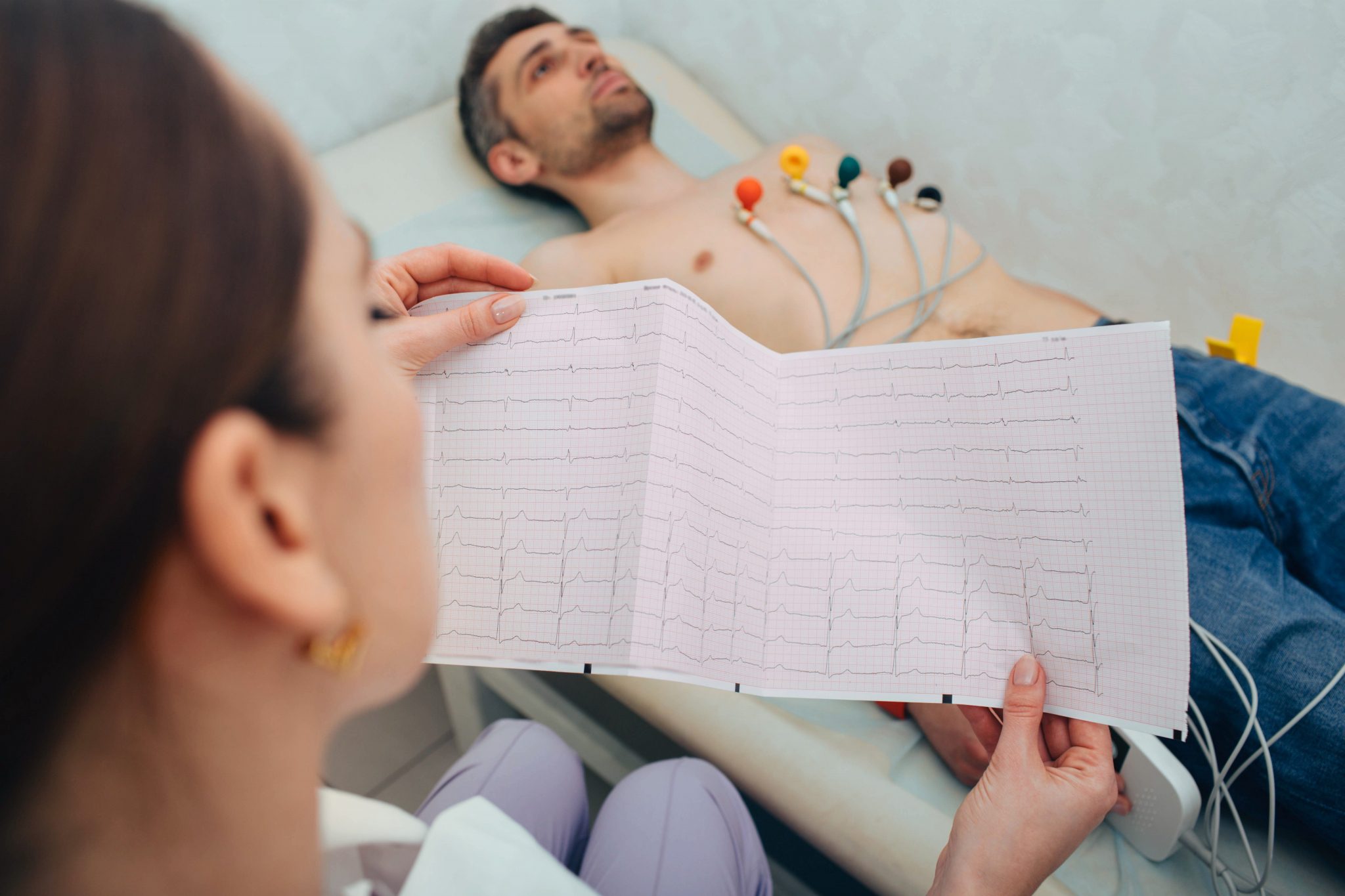
  1. Preparation: The patient typically lies down or reclines on an examination table or bed. Small adhesive electrodes, usually 10 in total, are attached to specific areas on the skin of the chest, arms, and legs. These electrodes act as sensors to detect the electrical signals generated by the heart.
  2. Recording: The ECG machine begins recording the electrical impulses as the heart beats. It measures the electrical activity along multiple pathways, creating a series of waves on a graph known as the ECG tracing.
  3. Interpretation: The ECG tracing is then interpreted by a trained healthcare provider, such as a cardiologist or electrocardiograph technician, who can identify any abnormalities or irregularities in the heart’s electrical pattern.

III. The Importance of ECGs in Healthcare

ECGs serve a vital role in healthcare for several reasons:

  1. Diagnosis: ECGs are instrumental in diagnosing a wide range of heart conditions, including arrhythmias (irregular heart rhythms), myocardial infarctions (heart attacks), and atrial fibrillation.
  2. Monitoring: ECGs are used to monitor patients with known heart conditions and to assess the effectiveness of treatments like medications or interventions such as pacemakers or defibrillators.
  3. Risk Assessment: They help assess an individual’s risk of heart disease and can be used as part of routine screenings or during preoperative evaluations.
  4. Research: ECG data plays a crucial role in heart-related research studies, helping researchers better understand heart conditions and treatment outcomes.

IV. Different Types of ECGs

While the standard 12-lead ECG is the most commonly used, there are other variations and specialized types of ECGs, including:

  1. Holter Monitor: This portable ECG device records heart activity over an extended period, typically 24 to 48 hours. It is often used to diagnose intermittent arrhythmias.
  2. Stress Test (Exercise ECG or Treadmill Test): During this test, the patient exercises on a treadmill or stationary bicycle while connected to an ECG machine. It helps assess heart function under stress and can uncover hidden cardiac issues.
  3. Ambulatory ECG (Event Monitor): This device records ECG data continuously over several weeks or months, allowing for long-term monitoring of heart activity. It is often used to detect infrequent arrhythmias or symptoms that occur irregularly.
  4. Signal-Averaged ECG: This specialized ECG focuses on assessing the electrical activity of the heart, particularly for patients at risk of ventricular arrhythmias or sudden cardiac death.

V. Interpreting ECG Results

Interpreting an ECG tracing requires training and expertise, but a basic understanding of the key components can help demystify the process:

  1. P Waves: These represent the electrical activity associated with atrial contraction. Abnormalities in the P waves can indicate atrial arrhythmias.
  2. QRS Complex: The QRS complex represents the electrical impulses that trigger ventricular contraction (the main pumping chambers of the heart). Changes in QRS duration or shape can signal ventricular arrhythmias or conduction abnormalities.
  3. T Waves: T waves correspond to the repolarization (relaxation) of the ventricles. Changes in T wave shape or amplitude can indicate various heart conditions, including myocardial ischemia or electrolyte imbalances.
  4. ST Segment: The ST segment is the flat, isoelectric section between the QRS complex and the T wave. Elevation or depression of the ST segment can be a sign of myocardial infarction (heart attack).
  5. QT Interval: The QT interval represents the time it takes for the ventricles to depolarize and then repolarize. Prolonged QT intervals can predispose individuals to life-threatening arrhythmias.

Electrocardiograms, or ECGs, are invaluable tools in cardiology and healthcare. They offer a non-invasive means of assessing the heart’s electrical activity, aiding in diagnosis, monitoring, and research. Understanding the basics of ECGs and their components can help patients and healthcare professionals communicate more effectively and make informed decisions about cardiac health. If you have concerns about your heart health or have been recommended for an ECG, it’s essential to consult with a qualified healthcare provider for a thorough evaluation and interpretation of the results.

“ECG” and “EKG” are two terms used interchangeably to refer to the same medical test, the electrocardiogram. However, there is a subtle difference in the terminology based on the language of origin:

  1. ECG (Electrocardiogram): “ECG” is the most widely used and internationally accepted abbreviation for the term. It originates from the German word “Elektrokardiogramm.”
  2. EKG (Electrokardiogram): “EKG” is primarily used in the United States. It is derived from the German word “Elektrokardiogramm” but uses the German spelling “Kardiogram” instead of the English “Cardiogram.”

In essence, both “ECG” and “EKG” refer to the same medical test that records the electrical activity of the heart. The choice of terminology may vary based on regional conventions and language preferences, but the underlying procedure and purpose remain identical.

What Does it Mean When My ECG is Abnormal

An abnormal electrocardiogram (ECG or EKG) means that the electrical activity of the heart, as recorded on the ECG tracing, deviates from the expected or normal patterns. It’s essential to understand that an abnormal ECG is not a diagnosis in itself; rather, it serves as a valuable tool for healthcare providers to identify potential heart-related issues that require further evaluation.

Here are some common reasons why an ECG may be considered abnormal:

Arrhythmias: Arrhythmias are irregular heart rhythms. An ECG can detect various types of arrhythmias, including atrial fibrillation (AFib), ventricular tachycardia (VT), bradycardia (slow heart rate), and others. Arrhythmias can lead to symptoms like palpitations, dizziness, or fainting.

Myocardial Ischemia: Abnormalities in the ECG, such as ST-segment depression or T-wave inversion, can indicate insufficient blood supply to the heart muscle. This condition, known as myocardial ischemia, may be a sign of coronary artery disease (CAD).

Myocardial Infarction (Heart Attack): A heart attack can result in specific ECG changes, such as ST-segment elevation or the development of pathological Q waves. These changes may indicate damage to the heart muscle due to reduced blood flow.

Conduction Abnormalities: ECG abnormalities can also reveal issues with the heart’s electrical conduction system, such as bundle branch blocks or heart block. These conditions can affect the heart’s ability to transmit electrical signals efficiently.

Hypertrophy: An abnormal ECG can suggest left ventricular hypertrophy (LVH) or right ventricular hypertrophy (RVH), which are conditions characterized by thickening of the heart muscle in response to increased workload.

Electrolyte Imbalances: Disturbances in blood electrolyte levels, such as low potassium (hypokalemia) or high calcium (hypercalcemia), can manifest as ECG abnormalities, potentially leading to arrhythmias.

Drugs and Medications: Certain medications, including some used to manage arrhythmias or hypertension, can cause ECG changes. It’s essential for healthcare providers to monitor ECGs in individuals taking such medications.

Structural Heart Abnormalities: ECG may reveal structural heart abnormalities such as atrial or ventricular septal defects, which are congenital heart conditions present from birth.

Pericarditis: Inflammation of the pericardium, the sac surrounding the heart, can lead to specific ECG changes, such as PR-segment depression or diffuse ST-segment elevations.

Non-Cardiac Factors: Sometimes, factors unrelated to the heart, such as muscle artifacts, improper lead placement, or patient movement, can lead to abnormal ECG readings. These are usually corrected with proper ECG technique.

It’s crucial to emphasize that an abnormal ECG alone does not provide a definitive diagnosis but serves as a valuable screening tool. Further evaluation, which may include additional tests like echocardiography, stress tests, or blood work, is typically needed to determine the underlying cause and appropriate treatment.

Patients with abnormal ECGs should consult with a healthcare provider, often a cardiologist, who can assess the ECG findings in the context of their medical history, symptoms, and other diagnostic tests. Timely evaluation and diagnosis are essential for appropriate management and the prevention of potential heart-related complications. One of the most common ECG (electrocardiogram) abnormalities seen in adults is atrial fibrillation (AFib). Atrial fibrillation is an arrhythmia characterized by irregular and often rapid heartbeats. In AFib, the atria (the upper chambers of the heart) quiver or fibrillate instead of contracting normally. This irregular electrical activity can lead to an irregular pulse and an ECG that shows erratic and irregular waves.

Key characteristics of atrial fibrillation on an ECG include:

  1. Absence of P Waves: Instead of clear, organized P waves representing atrial contraction, AFib typically shows chaotic and irregular fibrillatory waves.
  2. Irregular R-R Intervals: The distance between QRS complexes (R-R intervals) varies irregularly, indicating an irregular heart rate.
  3. Narrow QRS Complex: The ventricular response in AFib often results in a normal QRS complex duration, as long as the atrioventricular (AV) node conducts the irregular impulses appropriately.

Atrial fibrillation is a significant concern because it increases the risk of stroke due to blood pooling in the quivering atria, which can form clots that can travel to the brain. Additionally, AFib can lead to other complications, including heart failure and an increased risk of other arrhythmias. It’s important to note that while atrial fibrillation is one of the most common ECG abnormalities in adults, there are various other ECG findings and arrhythmias that healthcare providers may encounter, each with its own clinical significance and management approach. An accurate diagnosis and appropriate treatment are crucial for individuals with ECG abnormalities, and these should be determined by a healthcare professional, often a cardiologist, based on a thorough evaluation of the patient’s medical history, symptoms, and additional diagnostic tests.

FAQ About Your ECG

1. What is an ECG, and what does it measure?

Answer: An ECG, or electrocardiogram, is a medical test that measures the electrical activity of the heart. It records the heart’s electrical impulses as they travel through the heart muscle, producing a graphical representation of the heart’s rhythm and activity.

2. Why is an ECG performed?

Answer: ECGs are performed for various reasons, including:

  • Diagnosing heart conditions like arrhythmias, myocardial infarction (heart attack), and heart rhythm disorders.
  • Assessing the effectiveness of cardiac medications and interventions, such as pacemakers.
  • Evaluating the risk of heart disease in routine check-ups.
  • Monitoring heart health during surgical procedures or when patients are experiencing heart-related symptoms like chest pain, palpitations, or shortness of breath.

3. How is an ECG performed?

Answer: During an ECG, small adhesive electrodes are attached to specific areas on the skin of the chest, arms, and legs. These electrodes are connected to an ECG machine, which records the electrical signals produced by the heart as it beats. The test is painless and non-invasive, and the patient is typically required to lie still while the machine records the heart’s electrical activity.

4. What can an ECG reveal about heart health?

Answer: An ECG can provide valuable information about heart health, including:

  • The heart’s rhythm, helping to diagnose arrhythmias like atrial fibrillation or ventricular tachycardia.
  • Evidence of myocardial ischemia (reduced blood supply to the heart muscle), which can suggest coronary artery disease.
  • Signs of structural abnormalities in the heart’s chambers or walls.
  • The presence of conduction abnormalities, such as bundle branch blocks or heart blocks.
  • Changes that may indicate a previous heart attack or other cardiac conditions.

5. Is an abnormal ECG result a cause for concern?

Answer: An abnormal ECG result may indicate an underlying heart condition, but it doesn’t necessarily mean that a serious problem exists. Many factors can influence ECG readings, including age, medication use, and non-cardiac issues. Further evaluation by a healthcare provider, often a cardiologist, is essential to determine the cause and significance of the abnormality and to develop an appropriate treatment plan if necessary. Early detection and intervention can often lead to better outcomes for individuals with cardiac issues.